Category

Solutions

X-Ray Sources for Micro CT Systems

X-Ray Sources for Micro CT and Industrial CT Systems

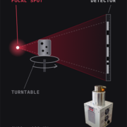
A micro CT scanner builds a 3D model from hundreds of X-ray pictures taken as a part rotates. The X-ray source sets the ceiling on how small a detail the finished scan can show. Scanner builders need focal spot size matched to their voxel, tube voltage set by the part’s material, and spot position stability that holds across a scan lasting hours. The CX20 carries the most flux in the range at 25 W. The CX30 reaches 130 kV and publishes its full spot against power curve. The CX40 holds the smallest spot at 4.5 μm.
Biopsy Specimen Radiography X-Ray Sources

X-Ray Sources for Medical and Biopsy Specimen Radiography

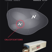
When a surgeon removes tissue or a radiologist takes a biopsy core, someone has to confirm the target came out before the patient leaves the room. A cabinet X-ray exposure shows the calcifications, the marker clip and the tissue edges in seconds. A lab micro-CT scan checks the margin from every angle afterward. Specimen size sets the power needed, from about 7 W for a block or core up to 50 W for a whole specimen. Tissue keeps the voltage low, between 20 kV and 55 kV. The CX40 covers that entire voltage band and holds a 4.5 μm focal spot for blocks and cores. The CX30 40 W model suits a whole specimen.

High Voltage Power Supply X-Ray Inspection

X-Ray Inspection of High-Voltage Power Supplies and Power Modules

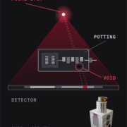
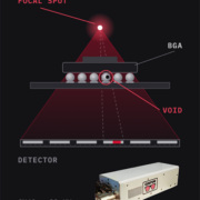
Manufacturers cast a high-voltage power supply in resin, so the resin fills every gap that would otherwise let voltage arc across it. Once the resin sets, nobody can open the unit to check it. X-ray finds voids in the potting, voids in the solder under a die, delamination, and parts that shifted during casting. Contrast decides most of this work, because a void in resin is a small density step inside an already light material. The CX30 covers the case, the copper windings and a power module’s copper substrate at 130 kV. Its 115 degree beam gives the off-angle views that separate a void from the layer holding it.

Counterfeit Electronics X-Ray Inspection | Curpow

X-Ray Detection of Counterfeit Electronic Components

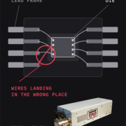
A counterfeit component carries the right part number on the outside and the wrong part inside. Opening it up to check destroys the component a screening lab needs to sell. X-ray shows whether a die exists at all, whether it matches a genuine component’s size, how many bond wires run inside the package, and where each one lands. MIL-STD-883 sets the published requirement at a 25.4 μm feature, which is the diameter of a signal bond wire. So resolution has to reach that scale. The CX40 reaches 4.5 μm and sits at the low end of the voltage range the method asks for.

Battery Cell X-Ray Inspection | Curpow

X-Ray Sources for Battery Cell Inspection

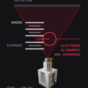
The last step of battery cell manufacturing is sealing. A sealed cell cannot be cut open without destroying the structure inside. Cell manufacturers use X-ray to confirm the anode covers the cathode edge, and to find metal particles in the stack. It also checks tab welds and confirms the electrolyte reached every layer. Pouch, prismatic and cylindrical cells each load the beam differently, and resolution needs to separate electrode layers about 20 μm apart. The CX20 names EV cell inspection directly on its data sheet. The CX30 handles in-line work and CT. The CX40 holds the smallest spot in the range.